Nutritional Aspects in Chronic Liver Disease

Disclaimer: This article is not intended as medical advice for patients and is for general information only.

This is an important but often overlooked area.

A lot of the guidelines on this are over 20 years old.

Malnutrition leads to multiple problems: piis1542356511011694_gr1_lrg

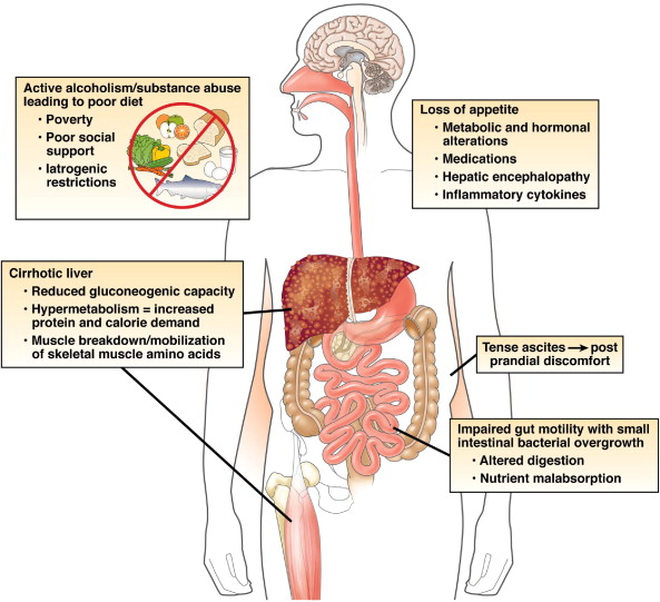
It is a vicious cycle. Particularly with the empty calories of alcohol.

There are often co-existant other medical problems such as: bile salt malabsorption, pancreatic exocrine insufficiency, SBBO, Neomycin.

This slideshow requires JavaScript.

The goal of treatment is to improve nutritional status and correct micro-nutrient deficiencies. Major options include oral/enteral supplementation or oral nasogastric/jejunal feeding.

All of the above are relatively simple measures to improve nutrition and they can have a dramatic influence on outcomes. In one study 1.Cabre et al patients with acute alcohol related hepatitis were randomised to steroids and nutrition or steroids alone. Only 8% of those who had nutrition plus steroids died, compared to 38% of those who only had steroids (p <0.05). The authors felt that the steroid only group had more infections, explaining the difference in mortality.

Other guidelines by 2. Plauth et al support this conclusion as well as the need for a low salt-diet in liver disease.

References:

  1. Cabré E, Rodríguez-Iglesias P, Caballería J, Quer JC, Sánchez-Lombraña JL, Parés A, Papo M, Planas R, Gassull MA. Short- and long-term outcome of severe alcohol-induced hepatitis treated with steroids or enteral nutrition: a multicenter randomized trial. Hepatology. 2000;32:36–42.
  2. Plauth M, Cabré E, Riggio O, Assis-Camilo M, Pirlich M, Kondrup J, Ferenci P, Holm E, Vom Dahl S, Müller MJ, et al. ESPEN Guidelines on Enteral Nutrition: Liver disease. Clin Nutr. 2006;25:285–294.

Cirrhosis and Pregnancy

Disclaimer: This article is not intended as medical advice for patients but is for informational purposes only.

liver-disease-in-pregnancy

Cirrhosis is when parts of the liver become hardened by damage done over time. Cirrhotic sections of liver unlike normal liver segments cannot repair itself.

Fortunately this is a relatively rare scenario as most patients develop cirrhosis later in life. However, cirrhosis is becoming more common due to rising rates of obesity. Pregnant women with cirrhosis  (which may be undiagnosed) can run into difficulty.

The biggest risk is bleeding, because pregnancy leads to an increased circulating volume of blood and the foetus compresses the inferior vena cava.

ivc-compression

Most of this bleeding occurs in the oesophagus and normally has to be prevented surgically either by banding of these vessels – called varices or inserting a shunt called a TIPS or transhepatic-portosystemicshunt. Pregnant women should ideally have foetal monitoring while undergoing any procedures.

This slideshow requires JavaScript.

Large liver-specialist centres need to be involved, especially if other problems subsequently develop. Early delivery may sometimes be necessary.

As long as the mother’s health is optimised then the same is normally true for the baby as in normal pregnancy.

Hepatitis

Hepatitis of any kind can be more serious in pregnancy. It needs to be managed with specialist input. Certainly chronic hepatitis B warrants some form of management at some point prior to birth in order to minimise the risk of transmission to the foetus.

Liver Imaging for Gastroenterologists

Disclaimer: This article is not intended as medical advice for patients but is for informational purposes only, primarily aimed at clinicians.

357127cf4b242ed836e6ad39dcd6b979

The liver is supplied by multiple vessels and divided into 8 segments.

In the normal liver most of the blood supply comes from the portal vein.

Tumours however tend to be supplied by the arterial supply (more on this later).

Regarding imaging. Ultrasound is great but unfortunately results are not reproducible between different operators.

o0006sup3dt1C

Common benign findings include: Cysts

liver_cyst_ultrasound

but sometimes they are not so simple

hyadtid
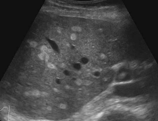
This one above turned out to be a hyatid cyst (Echinococcosis).

haemangioma

Haemangiomas can be even more difficult to distinguish.

focal-nodular-hyperplasia-i15

Focal nodular hyperplasia (above) can be distinguished from hepatomas by using Primavist (came out 2004). This contrast is taken up avidly by healthy hepatocytes and not by cancers.

Diffusion weighed MRI can help to distinguish between small metastases and other lesions, however its effectiveness is limited.

fatty-liver

Above is a fatty liver which makes it very difficult to distinguish the liver parenchyma

hepatitis

This is what acute hepatitis looks like on USS. The liver looks ‘brighter’.

The radiologist can also see vessels such as varices, abdominal collaterals, reversed/blocked portal flow and associated ascites etc.

Cirrhosis can be seen but only at an advanced stage so these imaging modalities are not so useful for the early detection of liver disease. Of CT, USS and MRI – MRI is the best at picking up cirrhosis but it is also the most expensive and difficult to access test.

Hepatocellular Carcinoma

The above conditions are all benign, but what we really want to spot are the cancers. These primary cancers normally occur in those with pre-existing liver disease.

Diagnosis can be very tricky if the timing goes wrong which is why you need a skilled radiology team.

This slideshow requires JavaScript.

Above are some images of HCCs. They tend to be surrounded by areas of necrosis as the tumour develops a rim of dead tissue around it.

Treatment options include:

This slideshow requires JavaScript.

Sorafenib, TACE, transarterial chemoembolization, resection and transplant

This is where we are now in 2016 but there are many other investigation and treatment options under investigation. So we can now tell what’s probably going on without doing a biopsy, most of the time. There is still significant margin for improvement.

Next up: Liver disease in pregnancy.