Urology and the medical registrar

Disclaimer: This article is meant for informational purposes only and not as clinical advice.

Warning: This article contains a few medical pictures

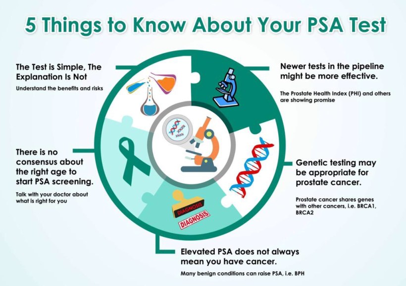
PSA / Prostate Cancer

img_1886

First discovered in the 1970’s. Produced in the prostatic ductal epithelial cells it is normally involved in the liquification of semen.

Prostate Cancer risk rises with age. As dose PSA. However, the thresholds for investigation are becoming lower and lower. Even a 30 year old, a PSA over 1 would warrant further investigation. Family history, recent instrumentation, racial origin, other genetic factors and drug/past medical history also all need to be taken into account.

PR exam, heavy cycling, BPH, urinary retention, prostatitis and UTI’s can all falsely elevate the PSA.

Older patients are less likely to have major surgery (prostatectomy etc). There is a 1% risk of sepsis following a prostate biopsy as well so this procedure has to be done with due consideration for the risks. There are newer tests such as MRI prostate and ‘template’ biopsies which are helping to get higher yields.

Anyone who has visible haematuria (blood in the urine) without an obvious cause needs to investigated. If the haematuria is invisible then there need to be two separate positive urine dips. In the major paper on this – Edwards et al, 2000. of almost 2,000 patients with visible haematuria 24% were found to have a urological malignancy.
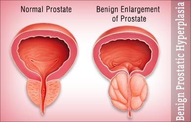
haematuria-dilutions-300x225
haematuria by dilution

BPH

BPH (Benign Prostatic Hypertrophy) is a histological diagnosis. The cause is not exactly known but it is to to do with adenomatous change in the prostate which pushes on and obstructs the ureter.

benign-enlargement-of-prostate

They will tend to have impaired flows and high post-void residual volumes on bladder scan. This doesn’t predict the likelihood of people going into retention but it does predict risk of needing treatment. If the post-void residual is >150mls then urologists recommend avoiding ‘anticholinergic’ medications.

Various treatment options for BPH include: alpha-blockers (these work over about 24 hours), 5ARIs – (dutasteride), anticholinergics, transurethral needle ablation, microwave thermotherapy.

Catheters

Some patients cannot be catheterised. If two tubes of instillagel fail try a Tiemann Tip catheter. In an emergency the bladder can be aspirated using a green needle transabdominally. Urologists however should be involved at this point. Irrigation stops future clots being formed. Washouts are needed to get rid of the clots. If this doesn’t work then they will need to go to theatre.

If in doubt call a urologist!

Neurology for the Non-Neurologist: Some unusual cases

Disclaimer: This article is intended as information only not medical advice. The cases are fictional.

gim-neurology
Neurons that fire together wire together

Rapid assessment in A&E:

Quick screening assessment can involve just getting the patient to walk, testing for vision and looking for tremors/weakness/abnormal movements as well as evident congnitive deficit. This + experience can help to rapidly distinguish between the ‘neurological’ emergency (stroke etc) and other differentials.

1. A case of unusual neurology: 60 year old man with tremor, abnormal gait and confusion. Gradual onset = Stroke is unlikely. ?Wernicke’s. Then his symptoms progressed including bilateral facial weakness and pain along right arm. None of this fits with a stroke. He then developed progressive opthalmoplegia (weakness of his eyes) and deteriorated further. MRI/CI – unhelpful. He had an lumbar puncture with extremely high white cells and protein. Then target like rashes were noted on his torso and spirochetes were found in the CSF. Diagnoses = Disseminated Neurological Lyme disease (rare). Treatment: Ceftriaxone 2 grams for 2 weeks. Other signs that can suggest Lyme: Conjunctivitis, Bilat weakness, positive serology, history of walking through forests / contact with certain animals.

Diagnosis: Neurological Lyme Disease

lyme
Tick bite with ring like skin rash

2. A fall with subsequent neurology: A 70 year old lady falls, hits her head and presents to A&E. On admission her chest x-ray looked normal. Initial CT of her head was normal. Then she becomes acutely short of breath and has a CT scan. This shows lots of blood in her chest (left side) – this is called a haemothorax. She has a chest drain inserted to help her breathing. The next day she is quite confused. ? Delirium (acute confusion related to painkillers/infection/trauma of admission). This was very out of character for her so a CT head was performed again. This looked relatively normal, however the brain looked large for someone in their 70’s (the brain shrinks with age), but was reported as normal (there was certainly no obvious abnormality). She then deteriorated and dropped her conscious level and had to be intubated. What is going on here? An LP was performed with white cell count of 1 and a protein level of 1.09. Neuroradiologists agreed that the CT scan looked normal. Then the PCR from the LP showed HSV type 1. MRI then confirmed this in the temporal lobes. This is a very dangerous condition that needs prompt and sometimes prolonged treatment with antiviral therapy.

Diagnosis: HSV Encephalitis Type 1

300px-hsv_encephalitis
HSV Type 1 Encephalitis affecting temporal lobes

3. Sudden unconsciousness ?cause. A  50 year old man who became suddenly unconscious (Glasgow Coma Score: 4/15). Examination was normal. He had to be intubated but all his other tests CXR, bloods, ECG were normal. He went on to have a CT scan of his head which was also normal. No evidence he had taken any drugs or toxins. Clinically infection didn’t appear likely. He had no evidence of vasculitis and there was no trauma. He had a second CT scan which showed an area of low density in the thalamus. MRI with DWI showed bilateral thalamic infacts.

Diagnosis: Thrombotic Stroke secondary to occlusion of a single dominant Artery of Percheron

annindianacadneurol_2016_19_3_426_179981_f1
Bilateral Thalamic Infarcts